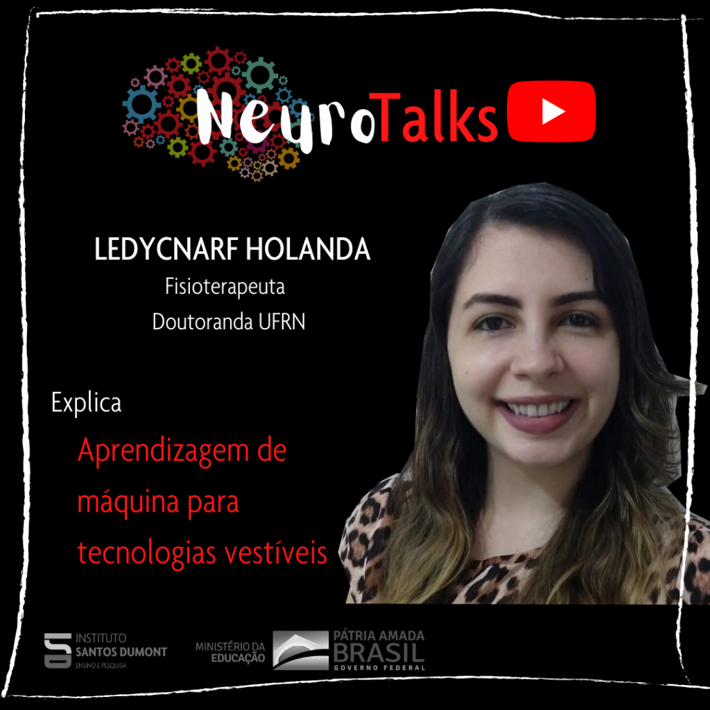
Ledycnarf Januário de Holanda1, Débora Cristina da Silva Oliveira2, Ana Paula Mendonça Fernandes2, Ricardo Alexsandro de Medeiros Valentim3, Danilo Alves Pinto Nagem3, Edgard Morya4, Ana Raquel Rodrigues Lindquist1,2
1Postgraduate Program in Physical Therapy, Department of Physical Therapy at the Federal University of Rio Grande do Norte, 2Graduation in Physical Therapy, Department of Physical Therapy at the Federal University of Rio Grande do Norte, 3 Department of Biomedical Engineering, Federal University of Rio Grande do Norte, 4Postgraduate Program in Neuroengineering, International Institute of Neurosciences – Edmond and Lily Safra, from Santos Dumont.
Summary
Machine learning (ML) is increasingly prevalent in technologies to offer a new perspective on how to live in society. One of the applications of AM has been in the improvement of tools for diagnosis and rehabilitation, through computational methods that use experiences acquired when exploring data to understand them and identify patterns, in order to improve performance and/or a predictive capacity. Thus, the evolution of technological devices in the areas of rehabilitation, telehealth and robotics enabled the development of more accurate and precise tools for monitoring physiological parameters during activities of daily living, thus, it is possible to stratify biological signals for better targeting of strategies therapies aimed at the health condition of each individual. Based on this, new technological resources have favored a better interaction between man and machine, providing the monitoring of biological signals in real time. In this context, there are wearable biofeedback systems able to continuously track the performance of a task and offer sensory stimuli, playing an important role in reducing the frequency of inappropriate movements, in order to detect and avoid inappropriate executions. These systems can be integrated with robotic devices, which have obtained promising results for improving sensorimotor function, functional capacity, and cardiorespiratory and metabolic conditioning. Above all, to improve mobility, quality of life, autonomy and awareness of health care, favoring the execution of tasks with greater efficiency and less physical overload, contributing to the functionality of people with motor dysfunctions. This favors the practice of therapies based on specific tasks, providing opportunities for the practice of preventive and rehabilitation measures, and increased adherence to more active therapies.
References
- HASSAN, Modar et al. Wearable gait measurement system with an instrumented cane for exoskeleton control. sensors, v. 14, no. 1, p. 1705-1722, 2014.2.
- HOLANDA, Ledycnarf J. et al. Robotic assisted gait as a tool for rehabilitation of individuals with spinal cord injury: a systematic review. Journal of neuroengineering and rehabilitation, v. 14, no. 1, p. 1-7, 2017.
- MENESES, Bárbara O. dos Santos et al. Innovation in assistive technology: opportunities and challenges. In: LEITE, Cicília Raquel Maia; REIS, Célia Aparecida dos; BINSFELD, Pedro Canisio; ROSA, Suélia de Siqueira Rodrigues Fleury (org.). New technologies applied to health: development of dynamic systems: concepts, applications and use of intelligent techniques and regulation. Mossoró – RN: EDUERN, 2019. E-book (608 p.). Available at: https://ppgcc.ufersa.edu.br/wp-content/uploads/sites/42/2019/07/novas-tecnologias-vol2-final3.pdf . Accessed on: December 13, 2020.
- SILVA, Patricia MM et al. Building Pressure-Sensitive Foot Insoles for Public Health Evaluation in Smart Cities. In: 2017 IEEE First Summer School on Smart Cities (S3C). IEEE, 2017. p. 153-156.
- STETTER, Bernd J. et al. A machine learning and wearable sensor based approach to estimate external knee flexion and adduction moments during various locomotion tasks. Frontiers in Bioengineering and Biotechnology, v. 8, 2020.
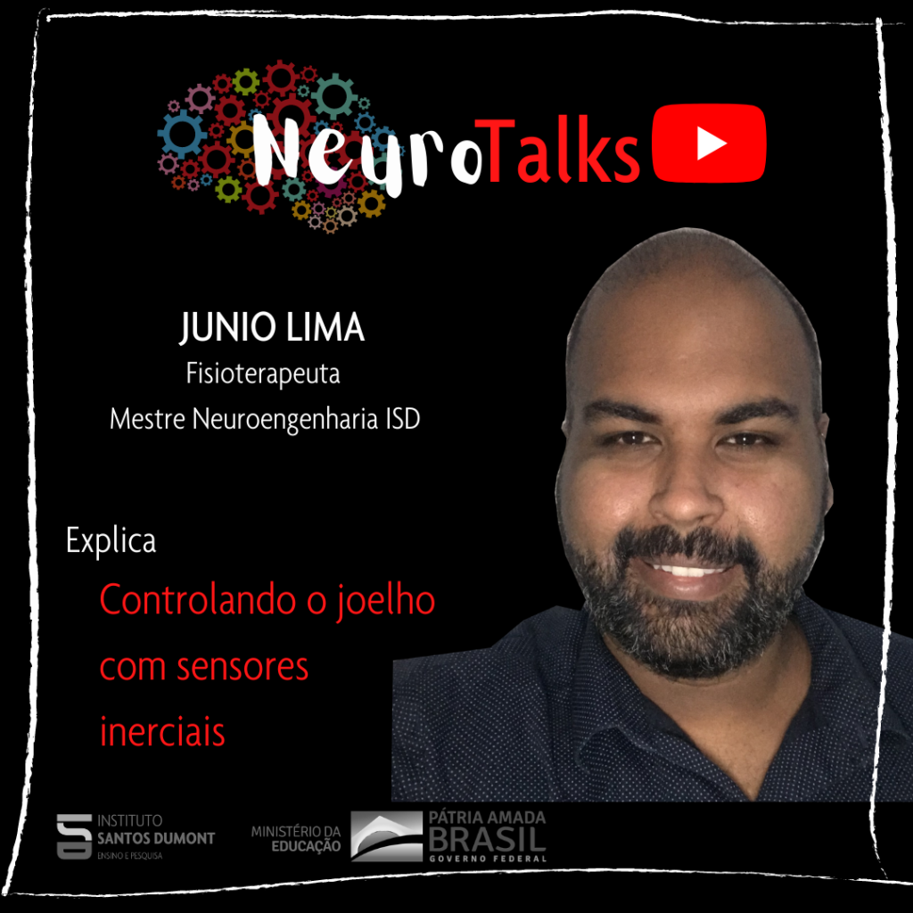
Junio Alves de Lima, Domingos Lira de Almeida Neto, Boaz Cavalcante Antunes Almeida, André Felipe Oliveira de Azevedo Dantas, Edgard Morya
Edmond and Lily Safra International Institute of Neurosciences, Santos Dumont Institute, Potiguar University, Laureate International Universities.
Summary
The development of systems that control and/or facilitate the functional movement of people with spinal cord injury, neurodegenerative diseases, stroke or other pathologies, are already a reality and object of study all over the world. Many devices use electrostimulation in their systems; however, the development of technologies that use electricity as their basis, requires adequate control in a multivariate way to execute movements similar to the physiological one for functional recovery. Therefore, the objective was to develop a functional electrical stimulation tool for knee movement using inertial information as feedback. The system is able to generate an alternating voltage ranging from 0-100 Hz (PW: 0-250μs; A: 100V; I: 100mA) and to calibrate in different impedances. A PID controller was implemented to determine the energy to be applied to the stimulated muscle to keep it in the pre-established position for 30 seconds. For system validation and testing, three points on the quadriceps were defined for stimulation. The controller was efficient to maintain the desired knee extension angle, even with external disturbances during its execution. Neurorehabilitation research and technological development enable real-time stimulation interfaces to integrate assistive technologies for functional recovery.
References
- KHAN, S.; QURESHI, R.; JAWAID, S.; SIDDIQUI, M.; SARWAR, U.; ABDULLAH, S.; KHAN, S.; KHAN, M.; BARI, A. Functional electrical stimulation (fes) based low-costassistive device for foot drop-a pilot study. In: SPRINGER.International Conference for Innovation in Biomedical Engineering and Life Sciences. [Sl], 2015.
- LAGUNA, Z.; CARDIEL, E.; GARAY, L.; HERNÁNDEZ, P. Electrical stimulator for surface nerve stimulation by using modulated pulses. In: IEEE.2011 Pan American HealthCare Exchanges. [Sl], 2011. p. 77–82.
- BOTTER, A.; OPRANDI, G.; LANFRANCO, F.; ALLASIA, S.; MAFFIULETTI, NA; MINETTO, MA Atlas of the muscle motor points for the lower limb: implications for electrical stimulation procedures and electrode positioning.European journal of applied physiology, Springer, v. 111, no. 10, p. 2461, 2011.
- JUNQUEIRA, MV; SANCHES, MA; KOZAN, RF; URBAN, MF; CARVALHO, AAD; TEIXEIRA, MC Development of an eight-channel functional electrostimulator for application with a feedback loop using a digital controller. 2013.
- SOETANTO, D.; KUO, C.-Y.; BABIC, D. Stabilization of human standing posture using functional neuromuscular stimulation.Journal of biomechanics, Elsevier, v. 34, no. 12, p.1589–1597, 2001.
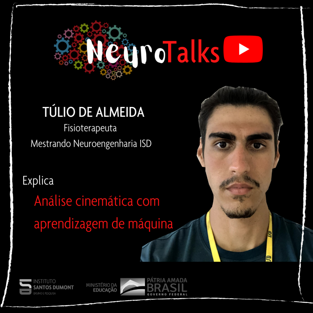
Túlio Fernandes de Almeida, Abner Cardoso Rodrigues Neto, André Felipe Oliveira de Azevedo Dantas.
International Institute of Neurosciences Edmond and Lily Safra.
Summary
Motor assessment, specifically gait, has been used for the characterization and early diagnosis of various diseases and disorders, such as Parkinson's disease. The use of inertial sensors and video analysis has been extensively researched in order to improve interventions for this purpose. One of the problems of using this technology is the acquisition price, the need for a highly controlled environment and extensive training for the use and interpretation of the data. In this context, the objective of the present study is to develop a wireless joint angle measurement system using inertial sensors. The main task to be evaluated in the present study is the human gait of healthy individuals through a protocol that involves other tasks, such as sitting down and climbing stairs. For the development of the work, 7 inertial sensors (MPU-6050) connected to 7 microcontrollers (ESP32) will be used through a 𝐼²𝐶 communication protocol. Data transmission will be performed using an Application Programming Interface through a Wi-Fi network. In order to avoid noise interference in the data, the performance of three filters will be used and compared: the Kalman Filter, the Complementary Filter and the MadgwickAHRS Filter. Finally, the system will also feature a software classifier using Principle Component Analysis and Suport Vector Machine to classify the tasks of moving from sitting to standing, going up and down stairs and each phase and subphases of normal gait.
References
- AN, K.-N. Kinematic analysis of human movement. Annals of biomedical engineering, Springer, v. 12, no. 6, p. 585–597, 1984.
- CHÈZE, L. Kinematic Analysis of Human Movement. [Sl]: John Wiley & Sons, 2014.
- HALILAJ, E.; RAJAGOPAL, A.; FITERAU, M.; HICKS, JL; HASTIE, TJ; DELP, SL Machine learning in human movement biomechanics: best practices, common pitfalls, and new opportunities. Journal of biomechanics, Elsevier, v. 81, p. 1–11, 2018.
- SEEL, T.; RAISCH, J.; SCHAUER, T. Imu-based joint angle measurement for gait analysis. Sensors, Multidisciplinary Digital Publishing Institute, v. 14, no. 4, p. 6891–6909, 2014.

Mirella Cunha Lira, Ramón Hypolito Lima
International Institute of Neurosciences Edmond and Lily Safra.
Summary
Since the description of the first brain rhythm, alpha waves, until the present day, knowledge about electrophysiological mechanisms and their implications has experienced great advances. Much as a result of the rise of increasingly precise techniques capable of detecting local oscillations, correlating them to resulting events and, finally, evaluating the oscillatory dynamics of neural circuitry, as happens in connectivity analyses. The range of available techniques makes it possible to visualize the same phenomenon from different perspectives, favoring discussions that are more faithful to what actually happens physiologically. The electroencephalogram (EEG), most used in clinical practice and therefore more popular, enabled many advances in the area, such as the study of evoked potentials and the diagnosis of epilepsy. The development of implantable multielectrode matrices allowed, therefore, the refinement of signal acquisition, attenuating the capture of noise and, due to proximity, capturing more preserved patterns, the local field potentials (LFP). action, the spikes. Even the investigation of the operation of ion channels of interest has become feasible thanks to the implementation of the patch clamp. Considering the relevance of oscillatory patterns for cognitive functions, the improvement of electrophysiological analysis tools brought new perspectives to the field of cognition. Thus, findings such as the characterization of oscillatory patterns conserved between individuals for specific tasks such as decision-making and memory consolidation, as well as electrophysiological signatures indicative of pathologies such as depression and epilepsy, support the construction of new diagnostic approaches and therapeutic perspectives innovations such as the use of electrostimulation.
References
- Buzsaki, G. (2006). Rhythms of the Brain. Oxford University Press.; Scanziani, M., & Häusser, M. (2009). Electrophysiology in the age of light. Nature, 461(7266), 930-939.
- Stevenson, WG, & Soejima, K. (2005). Recording techniques for clinical electrophysiology. Journal of cardiovascular electrophysiology, 16(9), 1017-1022.
- Sanchez, G., Daunizeau, J., Maby, E., Bertrand, O., Bompas, A., & Mattout, J. (2014). Toward a new application of real-time electrophysiology: online optimization of cognitive neurosciences hypothesis testing. Brain sciences, 4(1), 49-72.
- Pesaran, B., Vinck, M., Einevoll, GT, Sirota, A., Fries, P., Siegel, M., … & Srinivasan, R. (2018). Investigating large-scale brain dynamics using field potential recordings: analysis and interpretation. Nature neuroscience, 21(7), 903-919.

Valéria Azevedo de Almeida1, Edgard Morya1, Abner Cardoso Rodrigues Neto1, Lilian Lira Lisboa2, Rafael Pauletti Gonçalves2, Lucia Maria Costa Monteiro3, Reginaldo Antônio de Oliveira Freitas Júnior1.
1Edmond and Lily Safra International Institute of Neurosciences (IIN-ELS) Alberto Santos Dumont Teaching and Research Institute (ISD), RN 2Anita Garibaldi Health Education and Research Center (CEPS)- Alberto Santos Dumont Teaching and Research Institute (ISD) , RN 3National Institute of Women, Children and Adolescents Fernandes Figueira (IFF)- FIOCRUZ, RJ
There is scientific evidence that the Zika virus causes microcephaly and other neurological complications that together constitute Congenital Zika Virus Syndrome (CZS). Children exposed to fetal Zika virus infection present with structural cortical changes, spasticity, seizures, irritability, brainstem dysfunction, and neurogenic bladder. Understanding the neural mechanisms that modulate bladder activity in SCZ is a relevant factor in neuroscience and urology to improve therapeutic interventions. Simultaneous recording of bladder activity and neural activity in related brain regions makes it possible to advance in this still little explored field. In this perspective, the aim of this study was to investigate the relationship between voiding patterns and neural activity in children with SCZ. The research was approved by the Research Ethics Committee under registration CAAE-17583419.7.0000.5537. For the assessment of bladder function, the protocol published for the national cohort was used, established as recommended by the International Society of Child Continence. The urodynamic study was performed concurrently with the electroencephalogram. 10 children with confirmed SCZ, evaluated from July 2018 to December 2019, participated in this research. The extent of the impact of SCZ on the circuit between bladder and brain is still unknown and requires further research. There is evidence in animal models of alteration in Alpha and Theta frequencies during bladder overactivity, corroborating a possible modulation or alteration of the neural circuits involved in voiding control observed in children with CZS in this study. Considering the set of neurological manifestations involved in SCZ, further studies of cortical modulation and neurogenic bladder are needed to clarify the possible altered mechanisms.
References
- Chang HY, Havton LA (2008). Differential effects of urethane and isoflurane on external urethral sphincter electromyography and cystometry in rats. Am. J. Physiol. Renal Physiol. 295 F1248–F125.
- Costa monteiro, LM et al. Criteria to evaluate neurogenic bladder in children with Congenital Zika Syndrome. Protocols.io, dec. 2017.
- Costa monteiro, LM et al. Neurogenic bladder in the settings of Congenital Zika Syndrome: a confirmed and unknown condition for urologists. Journal of Pediatric Urology, Apr. 2019.
- Oliveira-Szejnfeld PS, Levine D, Melo ASO, et al. Congenital brain abnormalities and Zika virus: what the radiologist can expect to see prenatally and postnatally. Radiology. 2016;281:203-18.
- Yao, J., Li, Q., Li, X., Qin, H., Liang, S., Liao, X., & Yan, J. (2019). Simultaneous measurement of neuronal activity in the pontine micturition center and cystometry in freely moving mice. Frontiers in neuroscience.

Nancy Sotero Silva1, Carolina Karla de Souza Evangelista2, Danielly Carla da Silva Miranda1, Edgard Morya2
1Anita Garibaldi Health Education and Research Center, Santos Dumont Institute. 2Edmond and Lily Safra International Neuroscience Institute, Santos Dumont Institute.
Summary
Hearing losses can be caused by changes in structures from the outer, middle or inner ear (in sensitive and microscopic structures) to the auditory nerve. The various types of hearing loss have something in common: there is damage to the parts of the brain responsible for processing hearing information caused by the lack of sound stimulation. The sound, after being captured by the external ear, vibrates structures in the middle ear and is transformed into electrical information and sent by the auditory nerve to the auditory cortex, located in the temporal lobe of the brain. This region is divided into primary and secondary auditory cortex and also communicates with other regions of the brain, causing sound information to be interpreted as speech, for example, or as music, ambient sounds and noises. In addition, the auditory cortex has a true map, where each part is responsible for receiving and interpreting sounds at a specific frequency, thanks to a phenomenon called “tonotopy”. When a person has a hearing loss of only serious sounds, for example, the region of the auditory cortex responsible for processing this frequency range starts to process others. This process is called “neuroplasticity” and it is very important for people with hearing loss to be able to make the most of the sounds they can still hear. In children, this neuroplasticity is much greater, and so, when a child has a hearing loss and is rehabilitated with a hearing aid or cochlear implant, the regions of the auditory cortex that were occupied with other functions can do the opposite. A reorganization takes place so that the new sounds that they will now be able to hear are processed. In adults, however, few tests are performed to measure the effect of this neuroplasticity in auditory rehabilitation, and this investigation needs to become part of the clinical evaluation of speech therapy.
References
- DIETRICH, V.; NIESCHALK, M.; STOLL, W.; RAJAN, R.; PANTEV, C. Hearing Research, v. 58, pp. 95-101, 2001.
- EGGERMONT, JJ Hearing Research, v. 343, p. 176-190, 2017.
- KATZ, J. et al. (org.) Handbook of clinical audiology. Baltimore: Lippincott Williams & Wilkins, 2009.
- PEREZ, AP; ZILIOTTO, K.; PEREIRA, LD Test-retest of long latency auditory evoked potentials (P300) with pure tone and speech stimuli. International Archives of Otorhinolaryngology, v. 21, no. 2, p. 134–139, 1 Apr 2017.
- SHARMA, A.; DORMAN, MF; KRAL, A. The influence of a sensitive period on central auditory development in children with unilateral and bilateral cochlear implants. HearingResearch. v. 2013, no. 1-2. p 134-143, 2005.
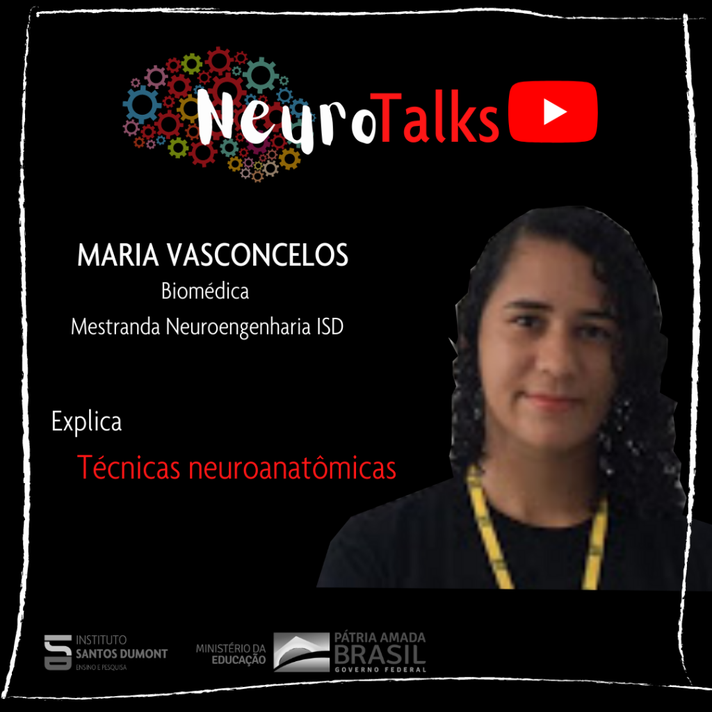
Maria Heloísa Moreira Vasconcelos and Carla Cristina Miranda de Castro.
Edmond and Lily Safra International Institute of Neurosciences – IIN-ELS
Summary
The purpose of this work is to present a synthesis of the main neuroanatomical methods used in research and their contributions, from the preparation of nervous tissue to techniques such as immunohistochemistry and immunofluorescence, aiming to answer questions related to neuroscience.
References
- PALETZKI, Ronald F.; GERFEN, Charles R. Basic Neuroanatomical Methods. Current protocols in neuroscience, v. 90, no. 1, p. e84, 2019.
- IF IMAGING: WIDEFIELD VERSUS CONFOCAL MICROSCOPY. ProteinTchec, 2019. Available at: https://www.ptglab.com/news/blog/if-imaging-widefield-versus-confocal-microscopy/. Accessed on: December 13, 2020.
- Nissl Body. Science direct, 2019. Available at: https://www.sciencedirect.com/topics/biochemistry-genetics-and-molecular-biology/nissl-body. accessed on December 13, 2020C
Valton da Silva Costa, Alice de Oliveira Barreto Suassuna.
International Institute of Neurosciences Edmond and Lily Safra.
Summary
We propose the realization of a video where intracortical microstimulation (MEIC) and medullary microstimulation (EME) will be approached. MEIC is an invasive technique for stimulation of the cerebral cortex, in which microelectrodes are surgically implanted in the nervous tissue, making it possible to inhibit or excite groups of neurons in different layers of the cortex. EME, on the other hand, is considered a semi-invasive technique, in which the electrodes are placed in the vertebral canal, over the spinal cord. The purpose of the video is to show the applications of microstimulation in neurosciences based on results obtained in recent experimental studies. It will present the effect of microstimulation on the primary somatosensory cortical area and adjacent areas, and the effects of medullary microstimulation on the microglial neuroinflammatory response, both from immunohistochemical analyzes in animal models. Based on these studies, the authors will discuss the implications of the results of these works in biomedical engineering and their applications in health, especially in neurology and neurorehabilitation.
References
- Ersen Ali, Elkabes Stella, Freedman David S, Sahin Mesut. Chronictissue response to untethered microelectrode implants in the rat brain and spinal cord. Journal of neural engineering. 2015;12:016019.
- Suassuna Alice de Oliveira Barreto, Silva Mayara Jully Costa, Oliveira João Rodrigo, et al. Microglial Activation After Acute Spinal Cord Electrode Implant in XXVI Brazilian Congress on Biomedical Engineering: 605–610 Springer 2019.
- O'Doherty Joseph E, Mikhail Lebedev, Timothy Hanson L, Nathan Fitzsimmons, Miguel Nicolelis AL. A brain-machine interface instructed by direct intracortical microstimulation. Frontiers in integrative neuroscience. 2009;3:20.
- Benali Alia, Weiler Elke, Benali Youssef, Dinse Hubert R, Eysel Ulf T. Excitation and inhibition jointly regulate cortical reorganization in adult rats. Journal of Neuroscience. 2008; 28:12284-12293.
Álisson de Oliveira Alves1, Edgard Morya1, Renan Cipriano Moioli2.
1Edmond and Lily Safra International Institute of Neurosciences, 2Federal University of Rio Grande do Norte.
Summary
Parkinson's disease (PD) is considered the second largest neurological disorder in the world, affecting about 1% of the world's population over 50 years of age. Clinically, this disease is characterized by muscle rigidity, resting tremor, bradykinesia and gait impairment, described as cardinal symptoms. At first, it is not possible to indicate a cure, however, the symptoms of the disease are relieved through the administration of chemical substances (such as Levodopa, currently the gold standard treatment) or through interventions that make use of electrical stimulation of deep brain regions or of the spinal cord. In this context, computational models emerge as a fundamental complement in the investigation of neurological disorders such as Parkinson's disease, facilitating the experimentation process, obtaining faster responses and reproducing phenomena that involve the functioning of the neural circuits involved in PD with great similarity. This work investigated the neural dynamics of brain regions involved in Parkinson's disease from dopamine depletion using a computational model of rats with 6-OHDA previously proposed in the literature. Using this model, it was possible to test and classify different patterns of the neural circuit involving the regions of the cortex – basal ganglia – thalamus – cortex through Principal Component Analysis (PCA) with simulations in three different states: healthy, parkinsonian, parkinsonian with cortical stimulus, thus simulating a stimulus coming from the motor cortex. The results of the work contribute to the development of interventions that may be more effective in suppressing the symptoms of PD, as a result of a more consolidated understanding of the mechanisms involved in the disease.
References
- HUMPHRIES, Mark D.; OBESO, Jose Angel; DREYER, Jakob Kisbye. Insights into Parkinson's disease from computational models of the basal ganglia. Journal of Neurology, Neurosurgery & Psychiatry, v. 89, no. 11, p. 1181-1188, 2018.
- KUMARAVELU, Karthik; BROCKER, David T.; GRILL, Warren M. A biophysical model of the cortex-basal ganglia-thalamus network in the 6-OHDA lesioned rat model of Parkinson's disease. Journal of computational neuroscience, v. 40, no. 2, p. 207-229, 2016.
- KANDEL, Eric et al. Principles of Neuroscience-5. AMGH Publisher, 2014.
- BEAR, MF; CONNORS, BW; PARADISO, MA Neurociencias: Unraveling the Nervous System, artmed R. Sao Paulo, Brazil, 2008.
- IZHIKEVICH, Eugene M. Simple model of spiking neurons. IEEE Transactions on Neural Networks, v. 14, no. 6, p. 1569-1572, 2003.
Luiz da Costa Nepomuceno Filho, Johseph Paballo Gomes de Souza, Maria Carolina Gonzalez, Ramon Hypolito Lima
Edmond and Lily Safra International Institute of Neuroscience IIN-ELS/ISD
Throughout its history, neuroscience has unraveled many mysteries about the functioning of the brain and nervous system. Many of these decades-long mysteries could be solved through the development of techniques that made it possible to understand not only how the brain works, but also how disorders affect it. One of these techniques is electrophysiology, in which researchers use arrays of microelectrodes, made with microwires smaller than a strand of hair, to record the neural signal while the animal is anesthetized or awake and moving freely . The manufacture of microelectrode arrays follows extremely precise stereotaxic orientations and coordinates and these are implanted in specific brain regions, in order to investigate what happens in these regions, making it possible to record the electrical activity of individual cells, groups of cells or interactions of large areas, according to the objective of the study. The electrode arrays can be purchased ready for implantation, which can cost more than a thousand dollars each, or they can also be manufactured, through specific methods of building microcircuits. The manipulation of these microcomponents is extremely delicate and requires a lot of training and accuracy, but the effort employed can promote great savings in the final price for each matrix. In this video we will discuss how the construction of microelectrode matrices is carried out, pointing out the main steps for their manufacture. Following the protocols developed by students and researchers at the International Institute of Neurosciences Edmond and Lily Safra, which enable investigations into the understanding of brain dynamics and the elucidation of great mysteries about the functioning of the brain and even the treatment of neurological diseases.
References
- BEAR, Mark; CONNORS, Barry; PARADISO, Michael A. Neuroscience: Exploring the brain. Jones & Bartlett Learning, LLC, 2020.
- MICROPROBES. OPTOGENETICS: OPTO-MICROELECTRODE ARRAY. In: Microprobes-Multi Channel Arrays Catalog, 2019. Available at: . Accessed on August 15, 2020.
- NICOLELIS, Miguel AL; LEHEW, Gary. Methods for Neural Ensemble recordings: Chapter 1State-of-the-Art Microwire Array Design for Chronic Neural Recordings in Behaving Animals. 2nd ed. Boca Raton: Crc Press/taylor & Francis, 2008. 11 p. Taylor & Francis Group, LLC.
- PAXINOS, George; FRANKLIN, Keith. Paxinos and Franklin's the Mouse Brain in Stereotaxic Coordinates. Gulf Professional Publishing, 2004. 360 p.
Paloma Cristina Alves de Oliveira, Thiago Anderson Brito de Araújo, Daniel Gomes da Silva Machado, Alexandre Hideki Okano, Suellen Mary Marinho dos Santos Andrade, Rodrigo Pegado de Abreu Freitas, Edgard Morya.
International Institute of Neurosciences Edmond and Lily Safra, International Institute of Neurosciences Edmond and Lily Safra, Federal University of Rio Grande do Norte, Federal University of ABC, Federal University of Paraíba, Federal Unit of Rio Grande do Norte, International Institute of Neurosciences Edmond and Lily Safra.
Summary
Parkinson's disease is a neurodegenerative disorder that causes motor and non-motor symptoms, being one of the main causes of disability in the world and the most growing neurological disease, affecting about 6.1 million people. Medications and neurosurgery are the main treatment options, however, they have disadvantages such as attenuation of effects after a few years of use, secondary complications, high cost or surgical risk. Because it presents a great challenge to science and health services, there is an urgent need for new therapies that are accessible and effective for the management of this disease. As a possibility, an innovative resource has aroused scientific interest for using a non-invasive, low-cost neuromodulatory technique, with minimal or no adverse effects and promising therapeutic potential. Transcranial Direct Current Stimulation is applied through a device with electrodes on the scalp, capable of modulating cognitive, motor and behavioral circuits. Its mechanism of action seems to include complex modes of plasticity, polarization of different neuronal regions, axonal growth, network effects and functioning of interneurons, endothelial and glial cells. Recent studies demonstrate that such a neuromodulatory approach may be able to improve several symptoms of Parkinson's disease, recently being classified as "probably effective". Indeed, this therapeutic modality seems to have the potential to provide a better quality of life for individuals and reduce the financial burden for society and health systems.
References
- FEIGIN, Valery L et al. Global, regional, and national burden of neurological disorders during 1990–2015: a systematic analysis for the global burden of disease study 2015. The Lancet Neurology, [SL], v. 16, no. 11, p. 877-897, Nov. 2017. Elsevier BV;
- FREGNI, Felipe et al. Evidence-based guidelines and secondary meta-analysis for the use of transcranial direct current stimulation (tDCS) in neurological and psychiatric disorders. International Journal Of Neuropsychopharmacology, [SL], p. 1-138, 26 Jul. 2020. Oxford University Press (OUP);
- JACKSON, Mark P. et al. Animal models of transcranial direct current stimulation: Methods and mechanisms. Clinical Neurophysiology, [sl], v. 127, no. 11, p.3425-3454, Nov. 2016. Elsevier BV;
- MORYA, Edgard et al. Beyond the target area: an integrative view of tdcs-induced motor cortex modulation in patients and athletes. Journal Of Neuroengineering And Rehabilitation, [SL], v. 16, no. 1, p. 1-29, 15 Nov. 2019. Springer Science and Business Media LLC;
- REICH, Stephen G.; SAVITT, Joseph M. Parkinson's Disease. Medical Clinics Of North America, [SL], v. 103, no. 2, p. 337-350, Mar. 2019. Elsevier BV.
Joao Paulo Bezerra Fernandes, Edgard Morya.
Edmond and Lily Safra International Institute of Neurosciences – IIN-ELS
Summary
Virtual reality (VR) is able to provide integration of people with disabilities into society. More than a billion people around the world live with a disability and can use VR resources to engage in their physiotherapeutic treatments, moving avatars in an immersive reality, the so-called exergames. A group that can exploit the technology's potential, but which still lacks structure, is formed by Paralympic boccia practitioners. Paralympic boccia is a sport played by athletes with cerebral palsy or neurological problems in which they use their hands to throw colored balls at a target ball, called a jack. Current training methods are based on indoor environments with little or no technology to assist coaches and athletes. Unlike other sports that rely on training high-tech athletes, bocce training doesn't have a lot of resources. iBoccia was developed to fill this gap and enable the use of an immersive reality that allows boccia athletes to train in a simulated environment and, at the same time, obtain data from their movements that will help in planning their training sessions under the supervision of coaches. To prove the effectiveness of the game, four experienced para-athletes of the modality evaluated iBoccia and, after a quantitative analysis of their performance, it was possible to evidence the improvement in the performance of the athlete's performance after using the platform. In this way, iBoccia stands as an innovative and unique game, whose purpose is to promote the modality and allow the improvement of the athletes' performance.
References
- Oh, Y., and Yang, S. (2010, November). Defining exergames exergam-ing. Proceedings of Meaningful Play, (pp. 1-17).
- Sherman, WR, Craig, AB (2018). Understanding virtual reality: Interface, application, and design.
- Morgan Kaufmann; Zhang, XW, Shyu, FM and You, GN (2018, April). The exergame for Tae-Bo learning with virtual reality. In 2018 IEEE International Conference on Applied System Invention (ICASI) (pp. 1018-1021). IEEE;
- Arndt, S., Perkis, A., and Voigt-Antons, JN (2018, October). Using Virtual Reality and Head-Mounted Displays to Increase Performance in Rowing Workouts. In Proceedings of the 1st International Workshop on Multimedia Content Analysis in Sports (pp. 45-50). ACM.
Victor Leandro da Cunha
International Institute of Neurosciences Edmond and Lily Safra.
Summary
Neural devices are widely used in neuroscience with the main objective of capturing neural signals that may bring answers about brain functioning and its connections. On the other hand, they can be used as microstimulators, acting in the modulation of specific regions for therapeutic purposes such as the treatment of Parkinson's disease and for scientific purposes in order to verify the neurophysiological and behavioral responses to certain stimuli (electrical, pharmacological or optogenetic). ). However, when implanted invasively in the brain tissue, it is possible to verify the loss of quality of electrophysiological recording and microstimulation in the long term. One of the possible causes that contribute to the reduction in the quality of recording and microstimulation is the neuroinflammatory response resulting from the process of penetration of the electrodes into the brain tissue, which causes an inflammatory response in which defense cells (microglia and astrocytes) will surround the implanted electrode in order to to combat the stressor agent, which ends up creating a barrier that hinders both the acquisition of signals and the distribution of microstimulation. Therefore, to reduce the glial encapsulation resulting from the neuroinflammation process, the insertion of electrodes is necessary, so that we can develop electrodes that are increasingly biocompatible with the tissue, in order to improve the quality of the acquired signal and the microstimulation offered when used in the long term. The present proposal aims to clarify the mechanisms involved in the neuroinflammatory response after the implantation of intracortical electrodes and to discuss both the physiological mechanisms and the technological challenges faced by scientists today. will pave the way for new studies to be carried out in this line of research.
References
- TSUI, C.; KOSS, K.; CHURCHWARD, M.; TODD, K. Biomaterials and glia: Progress on designs to modulate neuroinflammation. Acta biomaterialia, Elsevier, v. 83, p. 13–28, 2019.
- CHUNG, T.; WANG, J.; WANG, J.; CAO, B.; LI, Y.; PANG, S. Electrode modifications to lower electrode impedance and improve neural signal recording sensitivity. Journal of neural engineering, IOP Publishing, v. 12, no. 5, p. 056018, 2015.
- CHEN, R.; CANALES, A.; ANIKEEVA, P. Neural recording and modulation technologies. Nature Reviews Materials, Nature Publishing Group, v. 2, no. 2, p. 1–16, 2017.
- GULINO, M.; DONGHOON, K.; PANE, S.; SANTOS, SD; PÊGO, AP Tissue response to neural implants: the use of model systems towards new design solutions of implantable microelectrodes. Frontiers in neuroscience, Frontiers, v. 13, p. 689, 2019.
- SALATINO, JW; LUDWIG, KA; KOZAI, TD; PURCELL, EK Glial responses to implanted electrodes in the brain. Nature biomedical engineering, Nature Publishing Group, v. 1, no. 11, p. 862–877, 2017.

Carolina Karla de Souza Evangelista (1), Nancy Sotero Silva (2), Sheila Andreoli Balen (3), Edgard Morya (1).
(1) Edmond and Lily Safra International Neuroscience Institute, Santos Dumont Institute. (2) Anita Garibaldi Health Education and Research Center, Santos Dumont Institute. (3) Federal University of Rio Grande do Norte. Department of Phonoaudiology.
Summary
Identifying which newborns may develop a hearing and language disorder and intervening earlier is a challenge for public health. In the future, this may be possible with auditory evoked potentials (AEPs), which verify the response of components of the auditory pathway. Sometimes you can “hear” but you cannot “hear” or understand what is being said. This happens because other steps are involved in the act of “listening”. The way the brain analyzes and interprets sounds for us to understand is what we call auditory processing. It involves skills such as attention, sound discrimination, ability to understand speech in noise and sound localization. In addition, these skills are important for the development of oral and written language. For this to happen properly, all structures of the auditory system must be intact and the child must have access to quality auditory information. One way to verify these structures is to test with AEPs using electrodes on the scalp to capture the neural signals generated when we hear the sounds. There are short-latency AEPs, which assess from the auditory nerve to the brainstem nuclei, it is a procedure used to verify the integrity of the auditory pathway; and those of medium and long latency that present potentials originating in the primary and secondary auditory cortex and other areas such as the hippocampus, frontal cortex and thalamic areas. These last two still have poorly understood mechanisms with regard to their generating sites. Research that identifies the contributions of individual stages of auditory processing and develops systems for recording and processing the neural signal can serve as a tool to overcome this public health challenge.
References
- HALL, James W. Handbook of auditory evoked responses. Boston: Allyn & Bacon, 2006.
- HYPPOLITO, Miguel A. Evaluation of cortical auditory evoked potentials. In: Menezes et al (Org.), Treatise on Electrophysiology for Audiology. (pp. 97-116). São Paulo, SP: Booktoy, 2018.
- KRAUS, Nina; WHITE-SCHWOCH, Travis. Newborn Hearing Screening 2.0. The Hearing Journal, [SL], vol. 69, no. 11, p. 44-46, Nov. 2016.
- REGAÇONE, Simone F.; GUÇÃO, Ana CB; FRIZZO, Ana CF Electrophysiology: Current perspectives of its clinical application in speech therapy. Verba Volant, 2013.
- SCHOCHAT. Eliane. Middle Latency Auditory Evoked Potential. In: EDILENE MARCHINI BOÉCHAT (Org.). Textbook of Audiology. 2nd ed. Rio de Janeiro: Guanabara Koogan Ltda., 2015. Chap. 18. p. 135-139.
José Pablo Gonçalves de Queiroz, Livia Nascimento Rabelo, Fábio Henrique Medeiros Bezerra, Felipe Porto Fiuza.
Edmond and Lily Safra International Institute of Neurosciences (IIN-LES)/ Santos Dumont Institute (ISD)
The brain is responsible for coordinating all the body's functions, and it performs them in an integrated manner. Its operation depends on a dense and highly specialized architecture, which involves a complex system. Studying this dynamic entails a series of complex challenges. Thus, there are numerous approaches that can be used for its understanding. One of them is morphometry, which employs quantitative methods to measure structures, histological tissues, among others, not only applying to the field of neurosciences. Among the quantitative methods, the stereology technique is considered the gold standard for neuronal counting, since a rigorous sampling systematization methodology is employed in order to avoid bias, thus allowing the estimation of three-dimensional planes from two-dimensional planes, unlike of classical morphometry. In this perspective, the laboratory of quantitative neuroanatomy at the Santos Dumont Institute proposes to investigate morphometric parameters (glia/neuron ratio, neuronal and glial density) of histological images made available on an open access platform, exploring pathologies such as Alzheimer's, Vascular Accident and Disorder of the Autistic Spectrum. We used MBF Bioscience's Stereo Investigator software, version 11, to acquire and measure these parameters. Briefly, the histological images separated by sections are inserted into the program and then the anatomical region of interest is delimited. Afterwards, it is necessary to insert a grid, which basically works as a random event counting system and finally, manually and visually, the cells or structure of interest are identified using stereological markers. Its results make it possible to impact on premature diagnoses, prognoses, from a cellular characterization of different brain areas, contributing to scientific research and clinical results.
References
- ZHANG, Chen et al. A platform for stereological quantitative analysis of the brain-wide distribution of type-specific neurons. Scientific reports, v. 7, no. 1, p. 1-12, 2017.
- AGNATI, Luigi; FUXE, Kjell (Ed.). Quantitative neuroanatomy in transmitter research. Springer, 1985.
- CHERNIAK, Christopher. The bounded brain: toward quantitative neuroanatomy. Journal of Cognitive Neuroscience, v. 2, no. 1, p. 58-68, 1990.